v = dl/dt = rd(theta)/dt = r*omega
a = dv/dt = r*d(omega)/dt
= r*alpha
tau = F*r = mar = mr^2*alpha = I*alpha

2012년 11월 29일 목요일
Epoxides
II. Epoxides
Epoxides are compounds containing
the three-membered ring:

They are ethers, but the
three-membered ring gives them unusual properties which make them an exceedingly
important class of compounds. Epoxides are commonly made by the oxidation of
alkenes by peroxy compounds, such as benzoic acid:

When allowed to stand in ether or
chloroform solution, the peroxy acid and the unsaturated compound -- which need
not be a simple alkene -- react to yield benzoic acid and the epoxide. For
example:

Epoxides owe their importance to
the ease of opening of the highly strained three-membered ring. They undergo
acid-catalyzed reactions with extreme ease and -- unlike ordinary ethers -- can
even be cleaved by bases.
A polymer made of epoxide
units is called a polyepoxide or an
epoxy. Epoxy
resins are used as adhesives and structural materials; one such example is
epoxyethane.
Acid-catalyzed Cleavage
Like other ethers, an epoxide is
protonated by acid.

The protonated epoxide can then
undergo attack by any number of nucleophilic reagents.

An important feature of the
reactions of epoxides is the formation of compounds that contain two
functional groups. Thus, reaction with water yields a 1,2-diol. Reaction
with an alcohol yields a compound that is both ether and alcohol.
The two-stage process of
epoxidation followed by hydrolysis is stereoselective, and gives
1,2-diols corresponding to anti-addition to the C=C double bond.
The same stereochemistry was observed for hydroxylation of alkenes
by formic acid. There, an epoxide is formed as a reaction intermediate
which is rapidly cleaved in the acidic medium. The interpretation is
exactly the same as that given to account for anti-addition of halogens. Indeed,
epoxides and their hydrolysis served as a model on which the halonium ion
mechanism was patterned.
Base-catalyzed Cleavage
Unlike ordinary ethers, epoxides
can be cleaved under alkaline conditions. Here it is the epoxide itself -- not
the protonated epoxide -- which undergoes nucleophilic attack.

The lower reactivity of the
non-protonated epoxide is compensated for by the more basic, more strongly
nucleophilic reagents that are compatible with the alkaline solution (e.g.
alkoxides, phenoxides, ammonia, etc.).
Like alkyl halides and sulfonates,
and like carbonyl compounds, epoxides are an important source of
electrophilic carbon -- of carbon that is highly susceptible to attack by a
wide variety of nucleophiles. (E.G. Epoxides generated form carcinogenic
hydrocarbons are even attacked by the nucleophilic portion of the genetic
material DNA and thereby induce mutation and tumors).
Cleavage Orientation
There are tow C atoms in an
epoxide ring. In principle, either one can suffer nucleophilic attack. In a
symmetrical epoxide like ethylene oxide, the two carbons are equivalent, and
attack occurs randomly at either site. But in an asymmetrical epoxide molecule,
the C atoms are not equivalent, and the product obtained depends upon which one
is preferentially attacked.
It turns out that the preferred
point of attack depends chiefly on whether the reaction is acid-catalyzed or
base-catalyzed. Consider, for example, two reactions of isobutylene oxide:

Here (as in general) the
nucleophile attacks the more substituted carbon in an acid-catalyzed cleavage,
and the less substituted carbon in a base catalyzed cleavage.
Our first thought might be that
there are two different reaction mechanisms (e.g. SN1
vs. SN2).
But the evidence indicates clearly that both are of the SN2
type. This is characterized typically by cleavage of the C-O bond and attack by
the nucleophile in a single step.
How, then, are we to account
for the difference in orientation -- particularly for the SN2
attack at the more hindered position in acid-catalyzed cleavage ?
The answer to this query lies in
the transition state (or reaction intermediate).
In the transition state of
most SN2
reactions, bond-breaking and bond-making have proceeded to about the
same extent, and carbon has not become appreciably positive or negative.
Therefore steric factors, not electronic factors, chiefly
determine reactivity.
But in acid-catalyzed cleavage of
an epoxide, the C-O bond, already weak because of the angle strain
of the three-membered ring, is further weakened by protonation.
The leaving group is a very good one -- the weakly basic alcohol hydroxyl (OH)
group. Alternatively, the nucleophile is a poor one (e.g. water, alcohol). In
the transition state, bond-breaking has proceeded further than bond-making, and
thus carbon has acquired a considerable positive charge.

Since both leaving group and
nucleophile are far away, crowding is relatively unimportant here. The
stability of the transition state is determined chiefly by electronic factors
and not steric factors. Thus the reaction has considerable SN1
character. In this case:
Attack occurs at the C atom that can best accommodate the positive charge.
In base-catalyzed cleavage, the
leaving group is a poorer one -- a strongly basic alkoxide oxygen -- and the
nucleophile is a good one (e.g. hydroxide, alkoxide).

Bond-breaking and bond-making are
more nearly balanced, and reactivity is controlled in the more usual way
-- by steric factors. In this case:
Attack occurs at the less hindered carbon.
케톤의 알파-카본
Reactions at the α-Carbon
Many aldehydes and ketones undergo
substitution reactions at an alpha carbon, as shown in the following
diagram (alpha-carbon atoms are colored blue). These reactions are acid
or base catalyzed, but in the case of halogenation the reaction
generates an acid as one of the products, and is therefore
autocatalytic. If the alpha-carbon is a chiral center, as in the second
example, the products of halogenation and isotopic exchange are racemic.
Indeed, treatment of this ketone reactant with acid or base alone
serves to racemize it. Not all carbonyl compounds exhibit these
characteristics, the third ketone being an example.

Figure 1: Reactions at the  carbon
carbon
Two important conclusions may be drawn from these examples. First,
these substitutions are limited to carbon atoms alpha to the carbonyl
group. Cyclohexanone (the first ketone) has two alpha-carbons and four
potential substitutions (the alpha-hydrogens). Depending on the reaction
conditions, one or all four of these hydrogens may be substituted, but
none of the remaining six hydrogens on the ring react. The second ketone
confirms this fact, only the alpha-carbon undergoing substitution,
despite the presence of many other sites. Second, the
substitutions are limited to hydrogen atoms. This is demonstrated
convincingly by the third ketone, which is structurally similar to the
second but has no alpha-hydrogen.
Mechanism of Electrophilic α-Substitution
Kinetic studies
of these reactions provide additional information. The rates of
halogenation and isotope exchange are essentially the same (assuming
similar catalysts and concentrations), and are identical to the rate of
racemization for those reactants having chiral alpha-carbon units. At
low to moderate halogen concentrations, the rate of halogen substitution
is proportional (i.e. first order)
to aldehyde or ketone concentration, but independent of halogen
concentration. This suggests the existence of a common reaction
intermediate, formed in a slow (rate-determining step) prior to the
final substitution. Acid and base catalysts act to increase the rate at
which the common intermediate is formed, and their concentration also
influences the overall rate of substitution.
From previous knowledge and experience, we surmise that the common intermediate is an enol tautomer of the carbonyl reactant. Several facts support this proposal:
From previous knowledge and experience, we surmise that the common intermediate is an enol tautomer of the carbonyl reactant. Several facts support this proposal:
- Compounds that do not have any α-hydrogen atoms cannot enolize and do not undergo any of the reactions described above.
- The carbon-carbon double bond of an enol is planar, so any chirality that existed at the α carbon is lost on enolization. If chiral products are obtained from enol intermediates they will necessarily be racemic.
- In simple aldehydes and ketones enol tautomers are present in very low concentration. Reactions that involve enol reactants will therefore be limited in rate by the enol concentration. Increasing the amounts of other reactants will have little effect on the reaction rate.
- Enolization is catalyzed by acids and bases. These catalysts will therefore catalyze reactions proceeding via enol intermediates.
The reactions shown above, and others to
be described, may be characterized as an electrophilic attack on the
electron rich double bond of an enol tautomer. This resembles closely
the first step in the addition of acids and other electrophiles to alkenes.
Therefore, if electrophilic substitution reactions of this kind are to
take place it is necessary that nucleophilic character be established at
the alpha-carbon. A full description of the acid and base-catalyzed
keto-enol tautomerization process (shown below) discloses that only two
intermediate species satisfy this requirement. These are the enol tautomer itself and its conjugate base (common with that of the keto tautomer), usually referred to as an enolate anion.

Figure 2: Keto-enol tautomerization
Clearly, the proportion of enol tautomer
present at equilibrium is a critical factor in alpha substitution
reactions. In the case of simple aldehydes and ketones this is very
small, as noted above. A complementary property, the acidity of carbonyl
compounds is also important, since this influences the concentration of
the more nucleophilic enolate anion in a reaction system. Ketones such
as cyclohexanone are much more acidic than their parent hydrocarbons (by
at least 25 powers of ten); nevertheless they are still very weak acids
(pKa = 17 to 21) compared with water. Together with some
related acidities, this is listed in the following table. Even though
enol tautomers are about a million times more acidic than their keto
isomers, their low concentration makes this feature relatively
unimportant for many simple aldehydes and ketones.
Table 1: Acidity of α-Hydrogens in Some Activated Compounds
| Compound | RCH2–NO2 | RCH2–COR | RCH2–C≡N | RCH2–SO2R |
|---|---|---|---|---|
| pKa | 9 | 20 | 25 | 25 |
In cases where more than one activating
function influences a given set of alpha-hydrogens, the enol
concentration and acidity is increased. Examples of such doubly (and
higher) activated carbon acids are given elsewhere.

Tautomerism of aromatic compounds
Keto-Enol Tautomerism: Key Points

But every couple of blue moons (for acetone in water about 1/6600 th of the time at 23 °C) acetone undergoes a transformation to its alter ego, the enol form. [EDIT: equilibrium constant is 1 x 10^-8 in water) And as its name suggests, the enol form – which is an isomer, not a resonance form – has the characteristics of both alkenes and alcohols: it can involve itself in hydrogen bonding via the OH group, it is acidic at oxygen, and it reacts with electrophiles (like aldehydes, for example, in the Aldol reaction). In short, the enol form differs from the keto form in its polarity, acidity, and nucleophilicity just like werewolves differ from ordinary folks in their copious body hair, nocturnal rambunctiousness, and peculiar dietary habits.
The reason for the equilibrium lying to the left is due to bond energies. The keto form has a C–H, C–C, and C=O bond whereas the enol has a C=C, C–O an O–H bond. The sum of the first three is about 359 kcal/mol (1500 kJ/mol) and the second three is 347 kcal/mol (1452 kJ/mol). The keto form is therefore more thermodynamically stable by 12 kcal/mol (48 kJ/mol).
Although the keto form is most stable for aldehydes and ketones in most situations, there are several factors that will shift the equilibrium toward the enol form. The same factors that stabilize alkenes or alcohols will also stabilize the enol form. There are two strong factors and three subtle factors.
Biggies (2):
1. Aromaticity. Phenols can theoretically exist in their keto forms, but the enol form is greatly favored due to aromatic stabilization.
2. Hydrogen Bonding. Nearby hydrogen bond acceptors stabilize the enol form. When a Lewis basic group is nearby, the enol form is stabilized by internal hydrogen bonding.

Here are three more subtle effects in keto-enol tautomerism:
3. Solvent. Solvent can also play an important role in the relative stability of the enol form. For example, in benzene, the enol form of 2,4-pentanedione predominates in a 94:6 ratio over the keto form, whereas the numbers almost reverse completely in water. What’s going on? In a polar protic solvent like water, the lone pairs will be involved in hydrogen bonding with the solvent, making them less available to hydrogen bond with the enol form.
4. Conjugation . π systems are a little like Cheerios in milk: given the choice, they want to connect together than hang out in isolation. So in the molecule depicted, the more favorable tautomer will be the one on the left, where the double bond is a connected by conjugation to the phenyl.
5. Substitution. In the absence of steric factors, increasing substitution at carbon will stabilize the enol form. Enols are alkenes too – so any factors that stabilize alkenes, will stabilize enols as well. All else being equal, double bonds increase in thermodynamic stability as substitution is increased. So in the above example, the enol on the left should be the more stable one. As you might suspect, “all things being equal” sounds like a big caveat. It is - all else is rarely equal. But that’s a topic for another day – or, more likely, another course.

Sources: “March’s Advanced Organic Chemistry”, “Solvents and solvent effects in Organic Chemistry”, by Christian Riechart. EDIT: Commenter Natalia helpfully points out that Carey & Sundberg A is a great resource for this topic (section 7.3 in my 4th edition) and she is right.
2012년 11월 27일 화요일
spin-spin interaction

http://www2.chemistry.msu.edu/faculty/reusch/VirtTxtJml/Spectrpy/nmr/nmr1.htm
5.5A: The source of spin-spin coupling
The 1H-NMR spectra that we have seen so far (of methyl acetate and para-xylene)
are somewhat unusual in the sense that in both of these molecules, each
set of protons generates a single NMR signal. In fact, the 1H-NMR
spectra of most organic molecules contain proton signals that are
'split' into two or more sub-peaks. Rather than being a complication,
however, this splitting behavior actually provides us with more
information about our sample molecule.
Consider the spectrum for
1,1,2-trichloroethane. In this and in many spectra to follow, we show
enlargements of individual signals so that the signal splitting patterns
are recognizable.

The signal at 3.96 ppm, corresponding to the two Ha protons, is split into two subpeaks of equal height (and area) – this is referred to as a doublet. The Hb
signal at 5.76 ppm, on the other hand, is split into three sub-peaks,
with the middle peak higher than the two outside peaks - if we were to
integrate each subpeak, we would see that the area under the middle peak
is twice that of each of the outside peaks. This is called a triplet.
The source of signal splitting is a phenomenon called spin-spin coupling,
a term that describes the magnetic interactions between neighboring,
non-equivalent NMR-active nuclei. In our 1,1,2 trichloromethane example,
the Ha and Hb protons are spin-coupled to each other. Here's how it works, looking first at the Ha signal: in addition to being shielded by nearby valence electrons, each of the Ha protons is also influenced by the small magnetic field generated by Hb next door (remember, each spinning proton is like a tiny magnet). The magnetic moment of Hb will be aligned with B0
in (slightly more than) half of the molecules in the sample, while in
the remaining half of the molecules it will be opposed to B0. The Beff ‘felt’ by Ha is a slightly weaker if Hb is aligned against B0, or slightly stronger if Hb is aligned with B0. In other words, in half of the molecules Ha is shielded by Hb (thus the NMR signal is shifted slightly upfield) and in the other half Ha is deshielded by Hb (and the NMR signal shifted slightly downfield). What would otherwise be a single Ha
peak has been split into two sub-peaks (a doublet), one upfield and one
downfield of the original signal. These ideas an be illustrated by a splitting diagram, as shown below.

Now, let's think about the Hb signal. The magnetic environment experienced by Hb is influenced by the fields of both neighboring Ha protons, which we will call Ha1 and Ha2. There are four possibilities here, each of which is equally probable. First, the magnetic fields of both Ha1 and Ha2 could be aligned with B0, which would deshield Hb, shifting its NMR signal slightly downfield. Second, both the Ha1 and Ha2 magnetic fields could be aligned opposed to B0, which would shield Hb, shifting its resonance signal slightly upfield. Third and fourth, Ha1 could be with B0 and Ha2 opposed, or Ha1 opposed to B0 and Ha2 with B0. In each of the last two cases, the shielding effect of one Ha proton would cancel the deshielding effect of the other, and the chemical shift of Hb would be unchanged.

So in the end, the signal for Hb is a triplet, with the middle peak twice as large as the two outer peaks because there are two ways that Ha1 and Ha2 can cancel each other out.
Now, consider the spectrum for ethyl acetate:

We see an unsplit 'singlet' peak at 1.833 ppm that corresponds to the acetyl (Ha)
hydrogens – this is similar to the signal for the acetate hydrogens in
methyl acetate that we considered earlier. This signal is unsplit
because there are no adjacent hydrogens on the molecule. The signal at
1.055 ppm for the Hc hydrogens is split into a triplet by the two Hb
hydrogens next door. The explanation here is the same as the
explanation for the triplet peak we saw previously for
1,1,2-trichloroethane.
The Hb hydrogens give rise to a quartet signal
at 3.915 ppm – notice that the two middle peaks are taller then the two
outside peaks. This splitting pattern results from the spin-coupling
effect of the three Hc hydrogens next door, and can
be explained by an analysis similar to that which we used to explain the
doublet and triplet patterns.
| Example |
Exercise 5.6:
a) Explain, using left and right arrows to illustrate the possible combinations of nuclear spin states for the Hc hydrogens, why the Hb signal in ethyl acetate is split into a quartet.
b) The integration ratio of doublets is 1:1, and of triplets is 1:2:1. What is the integration ratio of the Hb quartet in ethyl acetate? (Hint – use the illustration that you drew in part a to answer this question.)
By now, you probably have recognized the pattern which is usually referred to as the n + 1 rule: if a set of hydrogens has n neighboring, non-equivalent hydrogens, it will be split into n + 1 subpeaks. Thus the two Hb hydrogens in ethyl acetate split the Hc signal into a triplet, and the three Hc hydrogens split the Hb
signal into a quartet. This is very useful information if we are
trying to determine the structure of an unknown molecule: if we see a
triplet signal, we know that the corresponding hydrogen or set of
hydrogens has two `neighbors`. When we begin to determine structures of
unknown compounds using 1H-NMR spectral data, it will become more apparent how this kind of information can be used.
Three important points need to be
emphasized here. First, signal splitting only occurs between
non-equivalent hydrogens – in other words, Ha1 in 1,1,2-trichloroethane is not split by Ha2, and vice-versa.

Second, splitting occurs primarily between hydrogens that are separated by three bonds. This is why the Ha hydrogens in ethyl acetate form a singlet– the nearest hydrogen neighbors are five bonds away, too far for coupling to occur.

Occasionally we will see four-bond and
even 5-bond splitting, but in these cases the magnetic influence of one
set of hydrogens on the other set is much more subtle than what we
typically see in three-bond splitting (more details about how we
quantify coupling interactions is provided in section 5.5B). Finally,
splitting is most noticeable with hydrogens bonded to carbon. Hydrogens
that are bonded to heteroatoms (alcohol or amino hydrogens, for
example) are coupled weakly - or not at all - to their neighbors. This
has to do with the fact that these protons exchange rapidly with solvent
or other sample molecules.
Below are a few more examples of chemical shift and splitting pattern information for some relatively simple organic molecules.


| Example |
Exercise 5.7: How many proton signals would you expect to see in the 1H-NMR
spectrum of triclosan (a common antimicrobial agent found in
detergents)? For each of the proton signals, predict the splitting
pattern. Assume that you see only 3-bond coupling.
Exercise 5.8: Predict the splitting pattern for the 1H-NMR
signals corresponding to the protons at the locations indicated by
arrows (the structure is that of the neurotransmitter serotonin).

5.5B: Coupling constants
Chemists quantify the spin-spin coupling effect using something called the coupling constant, which is abbreviated with the capital letter J.
The coupling constant is simply the difference, expressed in Hz,
between two adjacent sub-peaks in a split signal. For our doublet in
the 1,1,2-trichloroethane spectrum, for example, the two subpeaks are
separated by 6.1 Hz, and thus we write 3Ja-b = 6.1 Hz.

The superscript 3 tells us that this is a
three-bond coupling interaction, and the a-b subscript tells us that we
are talking about coupling between Ha and Hb. Unlike the chemical shift value, the coupling constant, expressed in Hz, is the same regardless of the applied field strength of the NMR magnet.
This is because the strength of the magnetic moment of a neighboring
proton, which is the source of the spin-spin coupling phenomenon, does not depend on the applied field strength.
When we look closely at the triplet
signal in 1,1,2-trichloroethane, we see that the coupling constant - the
`gap` between subpeaks - is 6.1 Hz, the same as for the doublet. This
is an important concept! The coupling constant 3Ja-b quantifies the magnetic interaction between the Ha and Hb hydrogen sets, and this interaction is of the same magnitude in either direction. In other words, Ha influences Hb to the same extent that Hb influences Ha. When looking at more complex NMR spectra, this idea of reciprocal coupling constants can be very helpful in identifying the coupling relationships between proton sets.
Coupling constants between proton sets on neighboring sp3-hybridized carbons is typically in the region of 6-8 Hz. With protons bound to sp2-hybridized
carbons, coupling constants can range from 0 Hz (no coupling at all) to
18 Hz, depending on the bonding arrangement.

For vinylic hydrogens in a trans configuration, we see coupling constants in the range of 3J = 11-18 Hz, while cis hydrogens couple in the 3J
= 6-15 Hz range. The 2-bond coupling between hydrogens bound to the
same alkene carbon (referred to as geminal hydrogens) is very fine,
generally 5 Hz or lower. Ortho hydrogens on a benzene ring couple at 6-10 Hz, while 4-bond coupling of up to 4 Hz is sometimes seen between meta hydrogens.

Fine (2-3 Hz) coupling is often seen between an aldehyde proton and a three-bond neighbor.
Table 4 lists typical constant values.
5.5C: Complex coupling
In all of the examples of spin-spin
coupling that we have seen so far, the observed splitting has resulted
from the coupling of one set of hydrogens to just one neighboring set of hydrogens. When a set of hydrogens is coupled to two or more sets of nonequivalent neighbors, the result is a phenomenon called complex coupling. A good illustration is provided by the 1H-NMR spectrum of methyl acrylate:

First, let's first consider the Hc signal, which is centered at 6.21 ppm. Here is a closer look:

With this enlargement, it becomes evident that the Hc signal is actually composed of four sub-peaks. Why is this? Hc is coupled to both Ha and Hb , but with two different coupling constants. Once again, a splitting diagram can help us to understand what we are seeing. Ha is trans to Hc across the double bond, and splits the Hc signal into a doublet with a coupling constant of 3Jac = 17.4 Hz. In addition, each of these Hc doublet sub-peaks is split again by Hb (geminal coupling) into two more doublets, each with a much smaller coupling constant of 2Jbc = 1.5 Hz.

The result of this `double splitting` is a pattern referred to as a doublet of doublets, abbreviated `dd`.
The signal for Ha at 5.95 ppm is also a doublet of doublets, with coupling constants 3Jac = 17.4 Hz and 3Jab = 6.31 Hz.

The signal for Hb at 5.64 ppm is split into a doublet by Ha, a cis coupling with 3Jab = 10.4 Hz. Each of the resulting sub-peaks is split again by Hc, with the same geminal coupling constant 2Jbc = 1.5 Hz that we saw previously when we looked at the Hc
signal. The overall result is again a doublet of doublets, this time
with the two `sub-doublets` spaced slightly closer due to the smaller
coupling constant for the cis interaction. Here is a blow-up of the actual Hb signal:

| Example |
Exercise 5.9: Construct a splitting diagram for the Hb signal in the 1H-NMR
spectrum of methyl acrylate. Show the chemical shift value for each
sub-peak, expressed in Hz (assume that the resonance frequency of TMS is
exactly 300 MHz).
When
constructing a splitting diagram to analyze complex coupling patterns,
it is usually easier to show the larger splitting first, followed by the
finer splitting (although the reverse would give the same end result).
When a proton is coupled to two
different neighboring proton sets with identical or very close coupling
constants, the splitting pattern that emerges often appears to follow
the simple `n + 1 rule` of non-complex splitting. In the spectrum of 1,1,3-trichloropropane, for example, we would expect the signal for Hb to be split into a triplet by Ha, and again into doublets by Hc, resulting in a 'triplet of doublets'.

Ha and Hc are not equivalent (their chemical shifts are different), but it turns out that 3Jab is very close to 3Jbc. If we perform a splitting diagram analysis for Hb,
we see that, due to the overlap of sub-peaks, the signal appears to be a
quartet, and for all intents and purposes follows the n + 1 rule.

For similar reasons, the Hc peak in the spectrum of 2-pentanone appears as a sextet, split by the five combined Hb and Hd protons. Technically, this 'sextet' could be considered to be a 'triplet of quartets' with overlapping sub-peaks.

| Example |
Exercise 5.10: What splitting pattern would you expect for the signal coresponding to Hb in the molecule below? Assume that Jab ~ Jbc. Draw a splitting diagram for this signal, and determine the relative integration values of each subpeak.

In
many cases, it is difficult to fully analyze a complex splitting
pattern. In the spectrum of toluene, for example, if we consider only
3-bond coupling we would expect the signal for Hb to be a doublet, Hd a triplet, and Hc a triplet.

In practice, however, all three aromatic
proton groups have very similar chemical shifts and their signals
overlap substantially, making such detailed analysis difficult. In this
case, we would refer to the aromatic part of the spectrum as a multiplet.
When we start trying to analyze complex
splitting patterns in larger molecules, we gain an appreciation for why
scientists are willing to pay large sums of money (hundreds of thousands
of dollars) for higher-field NMR instruments. Quite simply, the
stronger our magnet is, the more resolution we get in our spectrum. In a
100 MHz instrument (with a magnet of approximately 2.4 Tesla field
strength), the 12 ppm frequency 'window' in which we can observe proton
signals is 1200 Hz wide. In a 500 MHz (~12 Tesla) instrument, however,
the window is 6000 Hz - five times wider. In this sense, NMR
instruments are like digital cameras and HDTVs: better resolution means
more information and clearer pictures (and higher price tags!)
Contributors
- Organic Chemistry With a Biological Emphasis by Tim Soderberg (University of Minnesota, Morris)
This page viewed 13541 times
The ChemWiki has 7747 Modules.
Spin coupling in moleculesLooking at actual molecules raises questions about which nuclei can cause splitting to occur. First of all, it is important to realize that only nuclei with I ≠ 0 will show up in an NMR spectrum. When I = 0, there is only one possible spin state and obviously the nucleus cannot flip between states. Since the NMR signal is based on the absorption of radio frequency as a nucleus transitions from one spin state to another, I = 0 nuclei do not show up on NMR. In addition, they do not cause splitting of other NMR signals because they only have one possible magnetic moment. This simplifies NMR spectra, in particular of organic and organometallic compounds, greatly, since the majority of carbon atoms are 12C, which have I = 0.For a nucleus to cause splitting, it must be close enough to the nucleus being observed to affect its magnetic environment. The splitting technically occurs through bonds, not through space, so as a general rule, only nuclei separated by three or fewer bonds can split each other. However, even if a nucleus is close enough to another, it may not cause splitting. For splitting to occur, the nuclei must also be non-equivalent. To see how these factors affect real NMR spectra, consider the spectrum for chloroethane (Figure 4)Figure 4: The NMR spectrum for chloroethane. Adapted from A. M. Castillo, L. Patiny, and J. Wist. J. Magn. Reson., 2010, 209, 123.Figure 4 (Fig9.jpg)Notice that in Figure 4 there are two groups of peaks in the spectrum for chloroethane, a triplet and a quartet. These arise from the two different types of I ≠ 0 nuclei in the molecule, the protons on the methyl and methylene groups. The multiplet corresponding to the CH3 protons has a relative integration (peak area) of three (one for each proton) and is split by the two methylene protons (n = 2), which results in n + 1 peaks, i.e., 3 which is a triplet. The multiplet corresponding to the CH2 protons has an integration of two (one for each proton) and is split by the three methyl protons ((n = 3) which results in n + 1 peaks, i.e., 4 which is a quartet. Each group of nuclei splits the other, so in this way, they are coupled.
Coupling constantsThe difference (in Hz) between the peaks of a mulitplet is called the coupling constant. It is particular to the types of nuclei that give rise to the multiplet, and is independent of the field strength of the NMR instrument used. For this reason, the coupling constant is given in Hz, not ppm. The coupling constant for many common pairs of nuclei are known (Table 1), and this can help when interpreting spectra.Table 1: Typical coupling constants for various organic structural types.
Structural type
Coupling constant (Hz)
Fig10.jpg
6 - 8
Fig11.jpg
5 - 7
Fig12.jpg
2 - 12
Fig13.jpg
0.5 - 3
Fig14.jpg
12 - 15
Fig15.jpg
12 - 18
Fig16.jpg
7 - 12
Fig17.jpg
0.5 - 3
Fig18.jpg
3 - 11
Fig19.jpg
2 - 3
Fig20.jpg
ortho = 6 - 9; meta = 1 - 3; para = 0 - 1
Coupling constants are sometimes written nJ to denote the number of bonds (n) between the coupled nuclei. Alternatively, they are written as J(H-H) or JHH to indicate the coupling is between two hydrogen atoms. Thus, a coupling constant between a phosphorous atom and a hydrogen would be written as J(P-H) or JPH. Coupling constants are calculated empirically by measuring the distance between the peaks of a multiplet, and are expressed in Hz.Example 1Coupling constants may be calculated from spectra using frequency or chemical shift data. Consider the spectrum of chloroethane shown in Figure 5 and the frequency of the peaks (collected on a 60 MHz spectrometer) given in Table 2. Figure 5: 1H NMR spectrum of chloroethane. Peak positions for labeled peaks are given in Table 2. Figure 5 (Fig21.jpg)Table 2: Chemical shift in ppm and Hz for all peaks in the 1H NMR spectrum of chloroethane. Peak labels are given in Figure 5.
Peak label
δ (ppm)
ν (Hz)
a
3.7805
226.83
b
3.6628
219.77
c
3.5452
212.71
d
3.4275
205.65
e
1.3646
81.88
f
1.2470
74.82
g
1.1293
67.76
To determine the coupling constant for a multiplet (in this case, the quartet in Figure 5), the difference in frequency (ν) between each peak is calculated and the average of this value provides the coupling constant in Hz. For example using the data from Table 2:
Frequency of peak c - frequency of peak d = 212.71 Hz – 205.65 Hz = 7.06 HzFrequency of peak b - frequency of peak c = 219.77 Hz – 212.71 Hz = 7.06 HzFrequency of peak a - frequency of peak b = 226.83 Hz – 219.77 Hz = 7.06 HzAverage: 7.06 Hz
∴ J(H-H) = 7.06 HzNote: In this case the difference in frequency between each set of peaks is the same and therefore an average determination is not strictly necessary. In fact for 1st order spectra they should be the same. However, in some cases the peak picking programs used will result in small variations, and thus it is necessary to take the trouble to calculate a true average. To determine the coupling constant of the same multiplet using chemical shift data (δ), calculate the difference in ppm between each peak and average the values. Then multiply the chemical shift by the spectrometer field strength (in this case 60 MHz), in order to convert the value from ppm to Hz:Chemical shift of peak c - chemical shift of peak d = 3.5452 ppm – 3.4275 ppm = 0.1177 ppmChemical shift of peak b - chemical shift of peak c = 3.6628 ppm – 3.5452 ppm = 0.1176 ppmChemical shift of peak a - chemical shift of peak b = 3.7805 ppm – 3.6628 ppm = 0.1177 ppmAverage: 0.1176 ppmAverage difference in ppm x frequency of the NMR spectrometer = 0.1176 ppm x 60 MHz = 7.056 Hz∴ J(H-H) = 7.06 HzExercise 1
Calculate the coupling constant for triplet in the spectrum for chloroethane (Figure 5) using the data from Table 2.SolutionUsing frequency data:
Frequency of peak f - frequency of peak g = 74.82 Hz – 67.76 Hz = 7.06 Hz
Frequency of peak e - frequency of peak f = 81.88 Hz – 74.82 Hz = 7.06 Hz
Average = 7.06 Hz
∴ J(H-H) = 7.06 Hz
Alternatively, using chemical shift data:
Chemical shift of peak f - chemical shift of peak g = 1.2470 ppm – 1.1293 ppm = 0.1177 ppm
Chemical shift of peak e - chemical shift of peak f = 1.3646 ppm – 1.2470 ppm = 0.1176 ppm
Average = 0.11765 ppm
0.11765 ppm x 60 MHz = 7.059 Hz
∴ J(H-H) = 7.06 Hz
Notice the coupling constant for this multiplet is the same as that in the example. This is to be expected since the two multiplets are coupled with each other.
2012년 11월 26일 월요일
RING-OPENING AND RING-FORMING POLYMERIZATIONS
RING-OPENING AND RING-FORMING POLYMERIZATIONS:
A POLYMER SYNTHESIS AND CHARACTERIZAION EXPERIMENT
A POLYMER SYNTHESIS AND CHARACTERIZAION EXPERIMENT
INTRODUCTION:
Two experiments are actually combined into one through the concept of rings or cyclic structures, either in the monomer or the polymer formed. Seems a little tenuous, but these procedures are short and it does give you a different perspective on things. Background information is given on the general concepts of ring-opening and ring-forming polymerizations that includes industrial examples and mechanisms. The polymer synthesis for each type is combined with characterization by IR spectroscopy and dilute solution viscosity. The latter illustrates the polyelectrolyte effect for the charged ammonium polymer in water.
Polyamines, polyamine salts and polyamides are used commercially in a wide range of applications. These include sizing for paper and textiles; recovery and recycle of trace metal contaminates from chemical plants; and flocculation of particulate matter for water clarification. These polymers are strong complexing and chelating agents for metal salts, and such complexes have been used for a variety of catalytic applications. In addition, many of the polyamine compounds are basic catalysts in their own right or in conjunction with organic comonomers, and have been used to make a variety of synthetic chemicals.
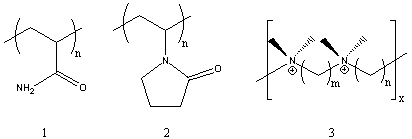
Several of the presently available commercial polymers containing amide and ammonium functionality are given in Figure 1. These included vinyl addition polymers from acrylamide (1) and N-vinylpyrrolidone (2), and step-growth polymers containing quaternary ammonium groups (polyionenes, 3) obtained from polycondensation of diamines and bishalides.
Figure 1. Commercial Amide and Ammonium Polymers. In this experiment, we examine two major types of polymerization processes involving heterocyclic monomers or repeat units. Polymer synthesis involves ring-opening polymerization to yield a polyamide from an oxazoline, and cyclopolymerization of diallylamine derivative to yield a polymer containing pyrrolidine units.
There are several commercially important polymers which are synthesized via ring-opening polymerization. Examples summarized in Figure 2 include such common polymers as polyoxyethylene (POE, 4), poly(butylene oxide) (PBO, 5), nylon 6 (6), and poly(ethyleneimine) (PEI, 7). This last polymer is obtained by a non-selective process which can involve attack on the ethyleneimine monomer by either chain-ends or internal secondary amines of the growing polymer. These competing reactions lead to a highly branched polymer structure which contains primary, secondary and tertiary amine units.1
Ring-opening polymerization mechanisms
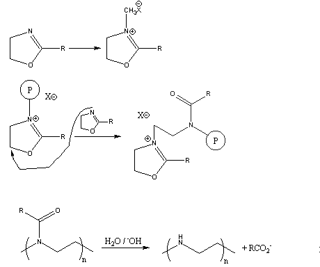
Figure 2. Common Ring-Opening Polymerizations. Several years ago, a novel synthesis of completely linear PEI was developed.2 The method utilized a ring-opening polymerization also, but of a 5-membered heterocycle that resulted in formation of a substituted amide rather than the free amine obtained from ethyleneimine. Figure 3 summarizes the initiation and propagation steps for this polymerization 3 as well as the hydrolysis reaction and the final polymer structure.
Figure 3. Ring-Opening Polymerization Mechanism for 2-Substituted Oxazolines
and Subsequent Polymer Hydrolysis; the Circled "P" Represents Polymer
Chain with the Indicated Active Chain End.
This method gives linear PEI (8) by a two-step process. In addition, the intermediate polymers containing amide functionality have become important in their own right.4 Many of these polymers are being examined for various unique applications involving a combination of properties. Many are soluble both in water and in a wide range of organic solvents. The amide functionality provides multiple sites for complexation and chelation of a variety of metal salts. The controlled spacing of the pendent amide derivatives along the 3-atom repeat unit in the backbone provides a novel alternative to the normal 2-atom backbone obtained with vinyl polymerization (see for example poly-(N-vinylpyrrolidone), 2). Finally, partial hydrolysis can give polymers containing both amide and amine or ammonium groups which can interact with substrates, together or in a sequential fashion.
The most common oxazoline derivative available today is the 2-ethyl compound. In this experiment, the monomer is polymerized using a cationic initiator to give high molecular weight polymer which is characterized by both IR and solubility behavior.
Cyclopolymerization
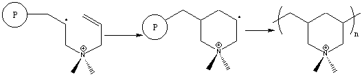
Cyclopolymerizations were first discovered by Professor George Butler in the late '50's.5 Since then, a wide variety of monomers have been found to undergo cyclopolymerization. We concentrate here on a diallylamine derivative. The cyclopolymerization process involves formation of a heterocyclic ring during polymerization as illustrated in Figure 4. The monomer shown, diallyldimethylammonium chloride, is one of the most widely used commercial derivatives.
The cyclopolymerization mechanism involves two sequential propagation steps.6 Intermolecular attack of a propagating radical is immediately followed by an intramolecular attack to form the heterocycle. Surprisingly, this second step leads to the unstable primary radical through kinetic rather than thermodynamic control, and is followed by immediate reaction with another monomer molecule. Two possible side reactions can occur in these polymerizations, involving crosslinking and chain transfer, but they are not observed. In general, cyclopolymerization of diallylammonium compounds proceeds cleanly to high molecular weight with no crosslinking.
Figure 4. Free Radical Cyclopolymerization Mechanism of a Diallylammonium Monomer
Figure 5. Free Radical Cyclopolymerization Alternate Mechanism of a Diallylammonium Monomer.
One of the earlier drawbacks in such polymerizations involved the use of peroxide initiators. Extensive yellowing of the product polymer and inefficient initiation lead to low yields and undesirable properties. A recently reported improvement on these polymerizations involves a new commercial initiator V-50 (2,2'-azobis(2-amidinopropane . 2HCl, 9). This water-soluble species cleanly forms carbon radicals that initiate diallylammonium cyclopolymerization to high yield.7
In this experiment, the polymerizability of diallyldimethylammonium chloride is examined. The polymer is purified by precipitation from water and characterized by dilute solution viscosity.
EXPERIMENTAL:
Polymerization of 2-Ethyloxazoline A clean, dry test tube is fitted with a rubber septum fastened on with wire. Approximately 2 ml of 2-ethyloxazoline8 is injected into the test tube which is then suspended in an oil bath preheated to 120oC. After a few minutes equilibration, the test tube is carefully removed and approximately 5ml of dimethylsulfate8 is injected. The test tube is put back in the oil bath. The solution gradually becomes more viscous until it gels or solidifies (about 2 h). The test tube is removed from the oil bath and allowed to cool. After removing the septum, 5 ml of methylene chloride is added to dissolve the mixture. This solution is then poured slowly into 50 ml of rapidly stirring mixed hexanes. The solvent is carefully decanted from the solid polymer which is washed again with more hexanes and finally isolated by filtration. CAUTION: dimethylsulfate is toxic and should be handled only in small quantities with good ventilation. Polymerization of Diallyldimethylammonium Chloride
Commercial monomer is usually available at 65 wt-% solution in water.9 This is suitable for direct polymerization. Approximately 5 ml of this solution is added to a test tube along with initiator 9 (V-509, about 0.05 g, ca. 1 mol-%). A septum cap is wired in place and the reaction mixture purged for 5-10 min with N2 through inlet and outlet needles in the septum. The test tube is placed in a preheated water or oil bath at 60-65oC. A small diameter syringe needle is left in the septum to relieve pressure from liberated N2 gas. Polymerization takes place rapidly to give a gelled or solid mass within 1-2 h. The polymer is isolated by precipitation into 100 ml ethanol stirring rapidly in a 250 ml beaker. Purification can be carried out by reprecipitation from water into ethanol.
DISCUSSION:
The two synthetic procedures are straight-forward and can be carried out with a minimum of special preparations and precautions. However, dimethylsulfate is toxic. Only enough material for immediate use should be used. The polymers are important commercially and represent less well-known specialty chemicals. In addition, their synthesis introduces the student to heterocyclic compounds in the context of polymer formation.
Two synthetic extensions of the experiment are possible. One involves synthesis of a poly(diallylamine)7, a polymer that is more difficult to purify and characterize. (Diallylamine is also toxic). Alternatively, the oxazoline polymer can by hydrolyzed in refluxing aqueous acid and neutralized to obtain the free amine polymer. These two polymers can then be compared with the other amide and amine polymers made in this experiment.
Polymer characterization involves qualitative evaluation of solubility behavior, dilute solution viscosity, and IR spectroscopy. Solubility should be evaluated for common organic solvents, acetic acid, and aqueous acid and base solutions. The results can be compared with other available polymers. The students must be made aware of the importance of allowing sufficient time for dissolution and swelling to take place (5-12 h). Unlike low molecular weight materials which normally dissolve rapidly or not at all, polymers take appreciable time to untangle and move away from the solid polymer mass. This effect becomes more pronounced the higher the molecular weight of the polymer.
Dilute solution viscosity is one of the most common and useful initial characterization techniques for polymers. At the very least, a viscosity value of more than ca. 0.1 dL/g tells you that you do have a polymer. More important, qualitative comparisons are possible for polymers of the same composition; ie., increasing viscosity values correlate directly with increasing molecular weight and polymer size. Detailed procedures have been published previously for viscosity determinations.
One very interesting aspect of the viscosity behavior of poly(diallyldimethylammonium chloride) is the polyelectrolyte effect that it shows.12 While well-behaved polymers show a linear relationship with respect to concentration, polyelectrolytes usually show higher reduced viscosity with decreasing concentration. This is demonstrated in the upper plot of Figure 6 (viscosity with units of dL/g plotted against concentration in g/dL). Addition of electrolytes (NaCl) at relatively high concentrations ( > 0.5M) compensates for the polyelectrolyte effect by masking the electrostatic repulsion of cationic groups along the polymer backbone. This is shown in the lower portion of Figure 6 where plots to two different types of viscosity values10,11 for the polymer plus electrolyte show linear behavior.
IR spectroscopy is the most routine spectral characterization technique available for polymers. The formation and IR characterization of polymer thin films is facile,13 giving both qualitative14 and quantitative15 information. Figure 7 gives the spectrum of the oxazoline polymer. Functional group identification can be required of the students, although polymer spectra often display unexpected combination bands and contaminant peaks from retained solvent and reactants.



Claisen Rearrangment
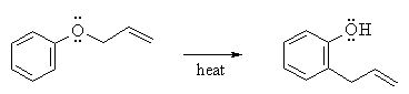
Claisen Rearrangment of Aryl Allyl Ethers
Reaction type : Electrocyclic reaction or sigmatropic rearrangement
Summary
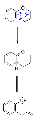
- Aryl allyl ethers undergo a thermal rearrangement to give ortho-allylphenols.
- This reaction is an intramolecular process.
- A sigmatropic rearangement is a reaction is which a σ bond migrates from one end of a π system to the other.
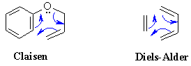
- Note how closely the mechanism of this reaction (table below) resembles that of the Diels-Alder reaction:
|
|
|
| Step 1:
An electrocyclic process. Push those electrons around the 6 membered ring.... Best appreciated by starting the electron flow by having the aromatic C=C attacking the allyl C=C and displacing an O leaving group.... |
 |
| Step 2:
Tautomerisation of the ketone (actually a dienone) to the more stable aromatic enol, the phenol. | |
피드 구독하기:
글 (Atom)